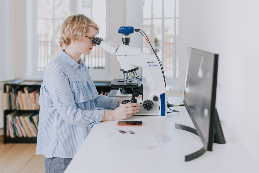
Cytology sample collection

This is a very concise overview of the most common errors encountered by VetCyto. I hope you find some helpful tips! Some of the tips are taken from the literature, some are based just on my personal experience, so take it with a “grain of salt”. If you have any comments or questions – please let us know

Cytology sample collection
Choose the method – FNA vs impression smear
- Both methods are good, but they are not the same! Clinician has to decide what questions to ask and what will be helpful.
- FNA – gives information about deeper tissues. This is a technique of choice if a mass is present/neoplasia is suspected.
- Impression smear – superficial material, frequently contains less informative material – secondary inflammation, cells from the skin surface etc. Good choice for seeing superficial pathology – infectious agents (malasezia, chailitiella etc).

Cytology sample collection
Aspiration vs Non-aspiration (capillary technique)
- Aspiration
– usually yields more cells
– better for poorly exfoliating masses (harder on palpation, lipomas)
– more traumatic to cells
– blood contamination more common
– be careful not to apply too much negative pressure in the syringe (~ 1cc is enough), move the plunger once and redirect the needle, repeat at least 4x
– always release negative pressure before the needle is taken out of the mass. This is important!
– once needle is out of the mass it is separated from the syringe
– 1 cc air is drawn in the syringe, needle is reattached and material is expelled on the slide
- Non-aspiration
– better for fragile cells (lymph nodes, endocrine tissues)
– can be performed with the needle only (syringe not attached)

Cytology sample collection
Redirect the needle
– Regardless if you aspirate or use non aspiration technique (capillary technique) – needle redirection is essential
– Redirection is movement of the needle/needle and syringe back and forth without taking it out of the mass
– Minimum 4 redirections are needed

Cytology sample collection
Needle size
– 22-25G needles are most commonly used. But every time after the palpation of the mass veterinarian has to choose the needle for that particular mass. The firmer/harder the mass – larger the needle (22G), softer mass – smaller needle diameter (23-25G).
– 22G needles can produce the core sample that is difficult to put on the slide in monolayer.

Slide preparation
– In my experience gentle rolling with the side (not the tip) of the needle you just used to obtain the aspirate is the best option
– Squash prep – squash the material between two slides is classically described in the text books, I find that this frequently causes too much pressure on the cells and they break
– Other methods can be used for liquid materials – more on sample preparation in the future.
References:
- Raskin RE, Meyer DJ. Canine and Feline Cytology A Color Atlas and Interpretation Guide. 3rd ed. Elsevier; 2016:1-15.
- Moore AR. Preparation of cytology samples: tricks of the trade. Vet Clin North Am Small Anim Pract. 2017;47

